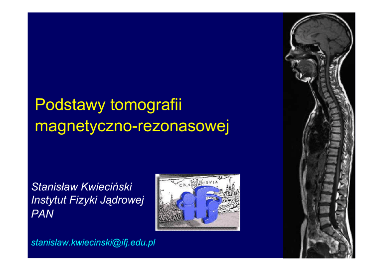
Podstawy tomografii
magnetyczno-rezonasowej
Stanisław Kwieciński
Instytut Fizyki Jądrowej
PAN
[email protected]
Optymistyczny plan
•
•
•
•
•
•
•
•
Pole magnetyczne
Spin jądrowy i moment magnetyczny
Magnetyzacja wypadkowa
Precesja
Magnetyczny Rezonans Jądrowy (MRJ)
Impuls RF
Czasy Relaksacji
Sygnał MR i jego parametry
•
•
•
•
Gradienty pola magnetycznego
Wybór warstwy
Kodowanie częstości
Kodowanie fazy
Sygnał
MR
Obraz
MR
Popularne techniki obrazowania
fotografia
zdjęcie Roentgenowskie
obraz MR
Jak otrzymać sygnał MR
obiekt
pole
magnetyczne B0
zmienne
pole B1
rezonans
surowy
sygnał
Transf.
Fouriera
„ładny”
sygnał
Magnes :
Cewka RF :
B0
B1
Cewki gradientowe: Gx, Gy, Gz
Cewka RF
Gradienty
Magnes
Tomograf MR
Magnetyczny Rezonans Jądrowy
MR
NMR
– Magnetic
– Nuclear
Resonance
Magnetic Resonance
– Rezonans(NMR)
Magnetyczny
Tomografia magnetyczno-rezonansowa
Tomografia – z jęz. greckiego
tomos - przekrój
grafo - rysuję
Obraz przekroju otrzymany przy wykorzystaniu
zjawiska magnetycznego rezonansu (jądrowego)
S
B1
Nuclear
B0
Signal
Magnetic
Resonace
N
Imaging
S
B1
Magnetyczny
B0
Sygnał MR
Rezonans
Jądrowy
N
Magnetic field
Some common examples
The Earth magnetic field
(B 0.05 T)
The magnetic field
lines of the bar magnet
Magnetic field
The magnetic field lines around
a long wire which carries
an electric current
The magnetic field lines
around circular loop through
which electric current flows
Różne rodzaje magnesów
1.5T, 3T, 7T
0.2-0.5T
Magnesy do zadań specjalnych
7, 9T
7 - 21T
Zadanie magnesu
Generowanie stałego pola magnetycznego Bo
Pole magnetyczne mierzymy w Teslach /T
Medyczne Tomografy MR maja pole 1.5T, 3T, 7T
Magnesy używane w laboratoriach generują pola nawet do 21T
W użyciu jest także stara jednostka pola Gauss / Gs
1T = 10000Gs
Pole ziemskie B 0.05 T
Nuclear spin J
Particle performing circular motion or rotating about the axis posses
angular momentum
Rotating nucleus has its own angular momentum called SPIN
NUCLEAR SPIN J (angular momentum of a nucleus)
J is a fundamental property of nature
J comes in multiples of 1/2 and can be + or –
(1/2, -1/2, 3/2, 5/2, -7/2…)
Protons, electrons, and neutrons have J = 1/2.
Nuclear spin
Nuclear magnetic moment
Nucleus is a charged object (due to protons) – rotating nucleus
generates a magnetic field
Nuclear magnetic moment μ
μ - nuclear
magnetic moment
γ - gyromagnetic
ratio
J – nuclear spin
μ =γ*J
Nuclear magnetic moment is
strongly associated with nuclear
spin although it is not the same
Nuclear magnetic moment μ
Question: Does every nucleus have a magnetic moment ?
Answer: No only these nuclei which have J > O
Pairs of protons (or neutrons) within a nucleus can cancel
each other out, so that only nuclei with an odd number of
either protons or neutrons will have a net magnetic moment
Hydrogen 1H
Lithium 7Li
Carbon 13C
Phosphorus 31P
1 proton
0 neutron
3 protons 4 neutrons
6 protons 7 neutrons
15 protons 16 neutrons
1
1
H
13
6
C
7
3
31
15
P
Li
Unfortunately nuclei with even number of protons and
neutrons have J=0 and therefore μ = 0
No magnetic moment means no chance for doing NMR /MRI
ie 12C, 16O, 56Fe
Magnetisation of a spin population
No net magnetization
Spin population in magnetic field
S
N
Magnetisation of a spin population
M
From a large
number of spins
We need only
consider the
difference that
makes the
majority
And this we can
represent as a
single vector M
(M -vector sum of all
spin magnetization
vectors in a volume
element)
Magnetisation of a spin population
B0
The ratio of induced magnetization M to
the applied field B0 is 10-9 10-10
M = B0
- magnetic susceptibility.
M
Magnetisation of a spin population
Spin energy in Magnetic Field
Energy
Spins antiparallel
higher energy
less stable
N+ / N- = exp (-E/kT)
Spins parallel
lower energy
more stable
Magnetic field strength B0
Jądro obdarzone momentem
magnetycznym oddziałowuje z
zewnętrznym polem magnetycznym Bo
Zjawisko Precesji
Precesja
B0
Zjawisko precesji spinu odkryte przez Larmora w 1895r.
0 = B0
M
B0 /T
Jądro
0.5
1
1.5
1
1
1H
1H
1H
13C
31P
Częstość
(MHz)
21
42
63
11
17
Rezonans
Częstość zewnętrzna (wymuszająca) = Częstości wewnętrznej systemu
fzewnętrzna = fwewnętrzna
Częstość zmiennego
pola magnetycznego
B1 (generowana
przez impuls RF)
1
B1
=
=
┴
Częstości precesji
(Larmora)
magnetyzacji M wokół
pola B0
0
B0
Przykład rezonansu
Katastrofa Mostu Tacoma
7 listopad 1940 r. USA
fwiatr = fmost
Gradient coil
Main Magnet
B0
RF Coil
Application of RF pulse
RF Coils
Generate the B1 field which excites the spins
to resonance and detect the resonance
signal.
To excite the resonance the B1 field must be
perpendicular to the main magnetic field B0.
Rodzaje cewek RF
(generujących pole B1)
powierzchniowe
Doty Scientific
macierze
NRC-IBD
objętościowe
GE
Philips
G. C. Wiggins et al. 2005
Cewka RF
typu „Bird Cage”
dla systemu 3T
Dzięki uprzejmości prof. B.Tomanka – NRC Canada
Gdań
Gdańsk 1 – 3.06.2006
ESMRMB School of MRI 2006
Czasy Relaksacji
RF Pulse
RF Excitation
Energy IN
z,B0
Mz
M
y
0
B1
x
Mx,y
M0
z,B0
T1
M
T2
x
0
y
Mx,y
Relaxation proceses (Recovery)
Transverse
Magnetization
Decays
Longitudinal
Magnetization
Recovers
Energy OUT
Detekcja sygnału MR
Zjawisko indukcji elektromagnetycznej
Surowy sygnał
(FID free induction decay)
z
x
y
Jak przetworzyć surowy sygnał
MR aby był bardziej czytelny?
Surowy sygnał MR (FID)
Transformata Fouriera
„Ładny” sygnał
Sygnał MR przed i po transformacie Fouriera
NMR Signal and its parameters
*
= 2/T2
0
Area under the curve, amplitude, are proportional to the number of
nuclei in the sample
The NMR signal width related to the transversal relaxation
Additional widening of the curve due to molecular motions, tunelling
effects , various couplings and interactions
Co możemy odczytać z sygnału MR ?
N
FIDCH3
FIDCH2
S
A liquid: C2H5OH
H
H
H C
C
H
H
OH
FIDOH
Spektroskopia MR etanolu CH3CH2OH
-CH3
-CH2 -
-OH
Gdań
Gdańsk 1 – 3.06.2006
ESMRMB School of MRI 2006
Jak otrzymać obraz MR?
O tym w części drugiej
Podziękowania
Prof. Andre JESMANOWICZ Medical College Wisconsin, USA
Dr. Christian KREMSER Innsbruck Medical University, Austria
Prof. Ludvikas KIMTYS Vilnius University, Lithuania
Prof.Andrzej Jasiński
Dr Tomasz Skórka
Dr Sylwia Heinze-Paluchowska
Mgr Krzysztof Jasiński
Mgr Anna Młynarczyk
Dr Katarzyna Majcher
Prof. Klaas Pruesmann
Dr Michael Wyss
Prof.Boguslaw Tomanek
Dr Wladyslaw Weglarz
Dr Tomasz Banasik
Dr Piotr Kulinowski
Dr Artur Krzyżak
Mgr Urszula Tyrankiewicz
Mgr Grzegorz Woźniak
Dziękuję
bardzo
za uwagę

