Opis przypadku/Case report
Endokrynologia Polska/Polish Journal of Endocrinology
Tom/Volume 63; Numer Number 4/2012
ISSN 0423–104X
Normal bone turnover markers in a patient
with active Paget’s disease of bone: response to treatment
with zoledronic acid
Prawidłowe stężenia markerów obrotu kostnego u pacjenta z aktywną chorobą
kości Pageta — odpowiedź na leczenie kwasem zoledronowym
Stergios A. Polyzos1, 2 Athanasios D. Anastasilakis3, Panagiotis Anagnostis2, Marina Kita2, Georgios Arsos4,
Efstratios Moralidis4, Athanasios Papatheodorou5, Evangelos Terpos6
Second Medical Clinic, Medical School, Aristotle University of Thessaloniki, Ippokration Hospital, Thessaloniki, Greece
Department of Endocrinology, Ippokration General Hospital, Thessaloniki, Greece
3
Department of Endocrinology, 424 General Military Hospital, Thessaloniki, Greece
4
Department of Nuclear Medicine, Aristotle University, Ippokration General Hospital, Thessaloniki, Greece
5
Department of Medical Research, 251 General Airforce Hospital, Athens, Greece
6
Department of Clinical Therapeutics, University of Athens School of Medicine, Athens, Greece
1
2
Abstract
The treatment of Paget’s disease of bone (PDB) aims at the suppression of abnormal bone turnover; bisphosphonates are currently the
treatment of choice. Indications for antiresorptive treatment in symptomatic patients with PDB include bone or joint pain, neurological
complications, surgery planned at an active pagetic site and hypercalcaemia from immobilisation. The goals of antiresorptive treatment
are clinical improvement and biochemical remission, as assessed by the normalisation of bone turnover markers. Clinical deterioration,
especially bone pain, should be considered before deciding to treat patients with late sclerotic (‘burned-out’) PDB. Bone scintigraphy may
be of importance in these patients, because it depicts increased osteoblastic activity, when bone markers may not. We present a case of
late sclerotic PDB with clinical deterioration but normal bone turnover markers, who experienced significant clinical improvement after
treatment with zoledronic acid. (Endokrynol Pol 2012; 63 (4): 312–315)
Key words: bisphosphonate, bone scintigraphy, bone-specific alkaline phosphatase, C-terminal telopeptide of type I collagen, Paget’s disease
of bone, zoledronic acid
Streszczenie
Celem leczenia choroby kości Pageta (PDB) jest zahamowanie zwiększonego obrotu kostnego. Obecnie lekami z wyboru są bisfosfoniany.
Do wskazań do stosowania leków antyresorpcyjnych u pacjentów z objawowa postacią PDB należą: bóle kostne i stawowe, powikłania
neurologiczne, planowany zabieg chirurgiczny w rejonie aktywnych zmian chorobowych i hiperkalcemia spowodowana unieruchomieniem. Celem terapii antyresorpcyjnej jest uzyskanie poprawy stanu klinicznego i remisji biochemicznej, ocenianej na podstawie
normalizacji stężeń biomarkerów obrotu kostnego. Przed podjęciem decyzji o wdrożeniu terapii u chorych w późnej, sklerotycznej fazie
choroby (burned out) należy wziąć pod uwagę pogorszenie stanu klinicznego, a zwłaszcza występowanie bólów kostnych. U tych chorych
duże znaczenie ma badanie scyntygraficzne kości, ponieważ może ono uwidocznić zwiększoną aktywność osteoblastyczną, której mogą
nie wykazać markery obrotu kostnego. W niniejszej pracy przedstawiono przypadek chorego w późnym, sklerotycznym stadium PDB,
u którego występowały nasilone objawy kliniczne, lecz stężenia markerów obrotu kostnego były prawidłowe. Po leczeniu kwasem zoledronowym nastąpiła istotna poprawa kliniczna. (Endokrynol Pol 2012; 63 (4): 312–315)
Słowa kluczowe: bisfosfoniany, scyntygrafia kości, izoenzym kostny fosfatazy alkalicznej, C-końcowy telopeptyd kolagenu typu I, choroba Pageta
kości, kwas zoledronowy
Introduction
Paget’s disease of bone (PDB) is the second commonest metabolic bone disease, after osteoporosis. PDB is
a chronic disorder characterised by focally high bone
remodelling, with abnormalities in all the phases of the
remodelling cycle, although the primary abnormality
is believed to lie in the regulation of osteoclasts, which
are increased in both number and size [1–3]. The treatment of PDB aims at the suppression of abnormal bone
turnover, and bisphosphonates (BPs) are currently the
treatment of choice [1, 4, 5]. Zoledronic acid (ZOL) leads
to quicker and more sustained therapeutic response
compared to other BPs such as risedronate [6, 7] or
Stergios A. Polyzos, 13 Simou Lianidi, 551 34 Thessaloniki, Greece, tel/fax: +30 231 042 47 10; e-mail: [email protected]
312
pamidronate [8]. Besides clinical improvement and biochemical remission, a favourable scintigraphic response
evident as early as three months after treatment with
ZOL has been reported in patients with PDB [9].
Indications for antiresorptive treatment in symptomatic patients with PDB include bone or joint pain,
neurological complications, surgery planned at an active
pagetic site and hypercalcaemia from immobilisation
[4, 5]. The goals of antiresorptive treatment are clinical
improvement and biochemical remission, as assessed by
the normalisation of bone turnover markers [10]. The
increased levels of bone markers are generally believed
to reflect the rate of bone remodelling and correlate
directly with the extent of the skeletal involvement
[11]. Total serum alkaline phosphatase (ALP) is the most
used marker in clinical practice, because it reflects the
disease activity and treatment efficacy and, additionally,
it is cheap, widely available, and has a low inter-assay
variability [4].
We hereby present a case of late sclerotic (‘burned-out’)
PDB with clinical deterioration but normal bone
turnover markers, who experienced significant clinical
improvement after treatment with ZOL.
roid hormone, and kidney and liver function tests were
within normal range.
In an attempt to deal with her clinical deterioration, the patient was administered a single 5 mg ZOL
infusion (October 2006), after a 10-day administration
of calcium and cholecalciferol supplements, as previously reported [9]. She did not experience an acute
phase reaction. She reported bone and articular pain
improvement, which started ten days after ZOL and
sustained for approximately 12 months. Notably,
she was able to walk without a stick from November
2006. As it has been elsewhere defined [4, 10], the
patient had an acceptable (> 25–30% decrease in
ALP) biochemical response to treatment at 6 months,
being a 33% decrease in ALP and a 36% decrease in
BALP, which lasted for at least 12 months and tended
to relapse at 18 months after ZOL (ALP: 86 IU/L at
6 months, 85 IU/L at 12 months, 113 IU/L at 18 months;
BALP: 25.2 IU/L, 27.9 IU/L, 44.5 IU/L, respectively).
However, serum CTX remained essentially unchanged (0.75 ng/mL, 0.66 ng/mL and 0.63 ng/mL,
respectively). The patient deceased from sudden
cardiac arrest in September 2008, being 85 years old.
Case description
Discussion
A Caucasian woman, born in 1923, was diagnosed with
symptomatic polyostotic PDB in 1980, (ALP 501, normal range 30–130 IU/L). She initially received courses
of calcitonin for 16 years (until 1996) with only partial
decreases in bone pain and ALP. Subsequently, when
ALP was increased, she had been receiving courses of
alendronate (1997 and 2001) and risedronate (2003 and
2005) treatment, which led to transient biochemical
and clinical remissions. The last course of risedronate
treatment was completed in March 2005. Since then,
the patient experienced progressively deteriorating
bone and articular pain, rendering her unable to walk
without the help of a stick from March 2006. Because of
clinical deterioration and the suspicion of malignancy,
she was subjected to bone scintigraphy (September
2006), which confirmed polyostotic PDB with enhanced
tracer uptake mainly in the skull, pelvis and spine, and
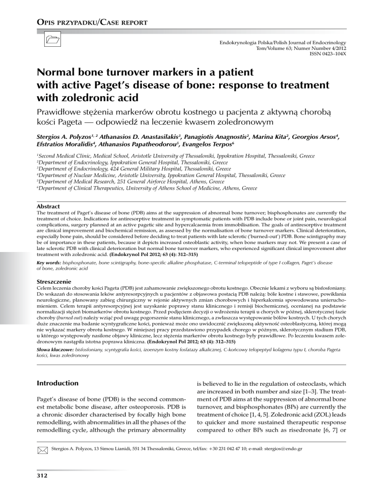
diffuse degenerative articular lesions (Figure 1A). Characteristically, plain radiographs were suggestive of late
sclerotic PDB (Figure 1B). Given that the low patient’s
ALP (129 IU/L) was incompatible with clinical deterioration, serum bone-specific alkaline phosphatase (BALP)
and C-terminal cross-linking telopeptide of type I collagen (CTX) were measured by established methods,
as elsewhere reported [12]; however, they were also
within normal range (BALP: 39.4 IU/L, normal range
14–43 IU/L; CTX: 0.66 ng/mL, normal range 0.12–
–0.75 ng/mL). Serum calcium, phosphate, albumin, parathy-
A woman with symptomatic, late sclerotic, polyostotic
PDB, who experienced significant clinical improvement
after treatment with ZOL, despite normal bone turnover
markers, is hereby presented.
There are limited similar cases in the literature. Ang
et al. described three patients with symptomatic active
PDB and normal ALP levels [13]. All three patients
had radiographic findings of PDB, increased uptake
of radiotracer on bone scintigraphy, but normal ALP;
however, they all had elevated levels of urinary markers of bone resorption. The patients were administered
pamidronate intravenously (60 mg once weekly for
2–3 consecutive weeks), after which bone pain and
scintigraphy were improved, urinary markers of bone
resorption were normalised, and ALP was decreased by
19–36% [13], approximately as in our case.
Gkouva et al. described a patient with symptomatic
monostotic PDB, but normal ALP and urinary hydroxyproline [14]. She had been treated for osteoporosis with
alendronate for the previous four years, which could
partly account for normal bone markers. Furthermore,
she had repeatedly received anti-inflammatory medication for tibia pain without significant relief. PDB was
confirmed by tibia plain radiograph, bone scintigraphy
and biopsy of the lesion. She was administered ZOL
intravenously (5 mg once), resulting in improvement
of bone pain and scintigraphy and a 45% decrease of
ALP [14].
313
OPIS PRZYPADKU
Endokrynologia Polska/Polish Journal of Endocrinology 2012; 63 (4)
Normal bone markers in Paget’s disease
Stergios A. Polyzos et al.
A
OPIS PRZYPADKU
B
Figure 1A. Bone scintigraphy showing polyostotic Paget’s disease of bone with enhanced tracer uptake more prominently on skull,
pelvis and spine, and dispersed degenerative articular lesions before zoledronic acid infusion; B. Radiograph of the skull showing
a ‘cotton-wool’ appearance indicative of the sclerotic phase of Paget’s disease of bone
Rycina 1A. Scyntygrafia kości przed podaniem we wlewie kwasu zoledronowego: cechy poliostotycznej choroby Pageta ze zwiększonym
wychwytem znacznika, zwłaszcza w kościach czaszki, miednicy i kręgosłupa, oraz rozsiane degeneracyjne zmiany stawowe; B. RTG
czaszki: zmiany typu „kłębków waty” sugerujące sklerotyczną fazę choroby kości Pageta
314
Endokrynologia Polska/Polish Journal of Endocrinology 2012; 63 (4)
In conclusion, active PDB may be present not only
with normal ALP levels, but also with normal BALP
and CTX. Clinical deterioration, especially bone pain,
should be considered before deciding to treat patients
with late sclerotic PDB. Bone scintigraphy may be
of importance in these patients, because it depicts
increased osteoblastic activity, when bone markers
may not. BPs treatment in these patients can lead to
clinical improvement.
Conflict of interest
There is no conflict of interest by any author pertinent
to this manuscript.
References
1.
2.
3.
4.
5.
6.
7.
8.
9.
10.
11.
12.
13.
14.
15.
16.
Polyzos SA, Anastasilakis AD, Terpos E. Paget’s disease of bone: emphasis
on treatment with zoledronic acid. Expert Rev Endocrinol Metab 2009;
4: 424–434.
Cundy T, Bolland M. Paget disease of bone. Trends Endocrinol Metab
2008; 19: 246–253.
Sheane BJ, Delaney H, Doran MF, Cunnane G. A classical presentation
of Paget disease of bone. J Clin Rheumatol 2008; 14: 373–373.
Devogelaer JP, Bergmann P, Body JJ et al. Management of patients with
Paget’s disease: a consensus document of the Belgian Bone Club. Osteoporos Int 2008; 19: 1109–1117.
Silverman SL. Paget disease of bone: therapeutic options. J Clin Rheumatol 2008; 14: 299–305.
Reid IR, Miller P, Lyles K et al. Comparison of a single infusion of zoledronic acid with risedronate for Paget’s disease. N Engl J Med 2005;
353: 898–908.
Hosking D, Lyles K, Brown JP et al. Long-term control of bone turnover
in Paget’s disease with zoledronic acid and risedronate. J Bone Miner
Res 2007; 22: 142–148.
Merlotti D, Gennari L, Martini G et al. Comparison of Different Intravenous Bisphosphonate Regimens for Paget’s Disease of Bone. J Bone
Miner Res 2007; 22: 1510–1517.
Avramidis A, Polyzos SA, Moralidis E et al. Scintigraphic, biochemical,
and clinical response to zoledronic acid treatment in patients with Paget’s
disease of bone. J Bone Miner Metab 2008; 26: 635–641.
Khan SA, McCloskey EV, Eyres KS et al. Comparison of three intravenous regimens of clodronate in Paget disease of bone. J Bone Miner Res
1996; 11: 178–182.
Falchetti A, Masi L, Brandi ML. Paget’s disease of bone: there’s more
than the affected skeletal-a clinical review and suggestions for the clinical
practice. Curr Opin Rheumatol 2010; 22: 410–423.
Polyzos SA, Anastasilakis AD, Litsas I et al. Profound hypocalcemia following effective response to zoledronic acid treatment in
a patient with juvenile Paget’s disease. J Bone Miner Metab 2010;
28: 706–712.
Ang G, Feiglin D, Moses AM. Symptomatic and scintigraphic improvement after intravenous pamidronate treatment of Paget’s disease of
bone in patients with normal serum alkaline phosphatase levels. Endocr
Pract 2003; 9: 280–283.
Gkouva L, Andrikoula M, Kontogeorgakos V, Papachristou DJ, Tsatsoulis
A. Active Paget’s disease of bone with normal biomarkers of bone
metabolism: a case report and review of the literature. Clin Rheumatol
2011; 30: 139–144.
Shankar S, Hosking DJ. Biochemical assessment of Paget’s disease of
bone. J Bone Miner Res 2006; 21 (Suppl 2): 22–27.
Garnero P, Christgau S, Delmas PD. The bisphosphonate zoledronate
decreases type II collagen breakdown in patients with Paget’s disease
of bone. Bone 2001; 28: 461–464.
315
OPIS PRZYPADKU
Unlike the Ang et al. cases [13], in our and Gkouva’s
patients [14], all measured markers were within normal
ranges. However, more sensitive bone formation and
resorption markers (BALP and CTX) were measured in
our case, which, in accordance to ALP, confirmed the
disease’s biochemical inactivity. Unlike the Gkouva case,
our patient had a long-established diagnosis of PDB,
which is consistent with her late sclerotic phase of PDB.
The decision for treatment was based on clinical and
scintigraphic criteria in all the above cases; the subsequent clinical and biochemical improvement supports
the necessity of treatment in these patients.
Although ALP is the bone marker of choice for the
diagnosis and follow-up of PDB patients [1, 4], BALP
is considered to be the most sensitive marker in the
evaluation of monostotic disease or in a case of limited
bone involvement, as it can be increased in up to 60% of
patients with normal ALP [1, 15]. However, even BALP
can be normal in very limited bone involvement [11].
ALP and BALP levels result from the increased number
of osteoblasts in the pagetic lesions. Their increased
levels reflect the rate of bone formation and correlate
directly with the extent of skeletal involvement [11].
During the natural course of PDB, ALP activity may
increase for many years, reflecting both expansion of
disease through affected bones and increased metabolic
activity [2]. After reaching its maximal extent and activity, the disease remains more or less stable for many
years, with relatively small fluctuations in ALP levels [2].
In accordance with this observation, it seems that
even the most sensitive markers, including BALP and
CTX, can be within normal ranges when PDB progresses
in the ‘burned-out’ phase, despite clinical deterioration.
Furthermore, given the unexpectedly distinct response
to ZOL of bone formation compared to bone resorption
markers, we speculate that CTX may not be a sensitive
marker for osteoclastic activity in cases of ‘burned-out’
PDB. The acceptable decrease in ALP, whose methods
are better standardised than CTX, is directly indicative of
a decrease in osteoblastic activity, and indirectly indicative
of both a decrease in osteoclastic activity and integrity
of the coupling mechanism (between osteoclastic and
osteoblastic activity) in our patient. The clinical improvement of our patient could be attributed to this response to
treatment, but also to a possible effect of ZOL on cartilage,
given that ZOL has been reported to decrease CTX-II,
thereby diminishing cartilage collagen degradation [16].

